EVAR (Endovascular Aneurysm Repair) is a minimally invasive procedure used to repair an abdominal aortic aneurysm (AAA) — a dangerous ballooning or weakening of the body's main artery, the aorta.
Instead of making a large abdominal incision like traditional open surgery, EVAR is performed from inside the blood vessel using catheters, guidewires, and a fabric-covered metal stent graft delivered through arteries in the groin.
EVAR
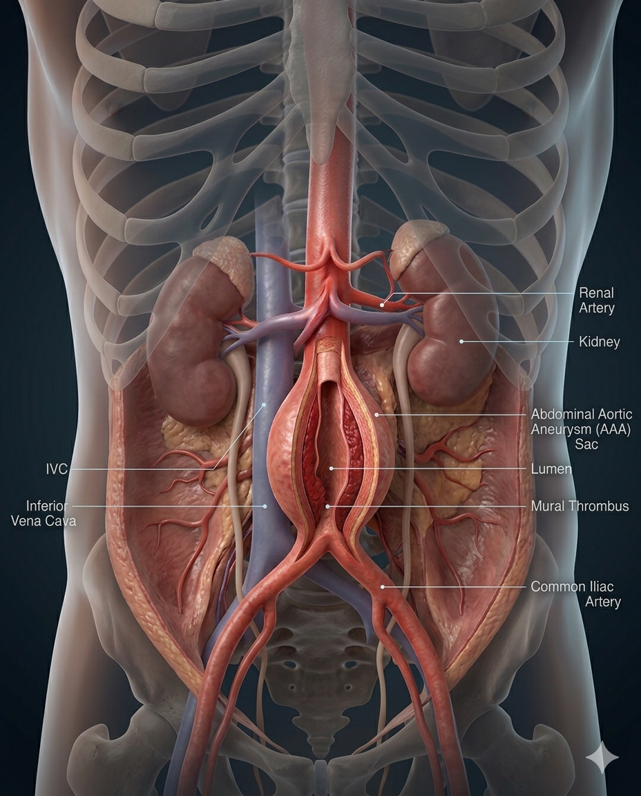
Abdominal Aortic Aneurysm (AAA) anatomy, transparent human torso, realistic abdominal cavity, infrarenal abdominal aorta enlarged into aneurysm sac, renal arteries, iliac arteries, inferior vena cava, kidneys, spine visible in background.
Cholecystectomy Digital Module
A guided, interactive, patient-to-professional digital experience that explains gallbladder disease, prepares the patient, maps the operation step by step, and connects anatomy, workflow, devices, risks, and recovery into one usable module.
A cholecystectomy is the surgical removal of the gallbladder, most commonly done laparoscopically for symptomatic gallstones, cholecystitis, or related biliary disease. Laparoscopic surgery is the usual approach, while open surgery may be needed in more difficult or converted cases. Safe identification of the cystic duct and cystic artery using the Critical View of Safety is a core principle in modern cholecystectomy.
-
This module should do four things at once:
1. Educate the patient in plain language
2. Support the surgeon and care team with structured workflow logic
3. Map devices, supplies, and pharma to each phase of the case
4. Create a reusable LDS digital product for web, app, LMS, and sales enablement.
-
A. Module title
Cholecystectomy: A Guided Digital Surgical Experience
B. Module audience
• Patients and families
• Surgeons
• OR staff
• Hospitals
• Device reps
• Pharma partners
• Educators and training programs
-
Patient-Facing Version
What is an abdominal aortic aneurysm (AAA)?
What is EVAR?
Why surgery may be needed
How EVAR repairs the aneurysm from inside the blood vessel
EVAR vs Open AAA Repair
Benefits, risks, and expected outcomes
Professional-Facing Version
AAA pathology
EVAR indications
Patient selection
Endograft principles
Anatomical considerations
• • Evidence and outcomes
-
Patient Layer
Aorta explained
Abdominal aorta location
Iliac arteries
Blood flow to organs and legs
What an aneurysm is
Professional Layer
Infrarenal aorta
Juxtarenal anatomy
Renal arteries
Iliac anatomy
Neck morphology
Landing zones
Tortuosity
Calcification
Access vessel anatomy
Visual Layer
3D abdominal aorta
Interactive aneurysm expansion model
• • Branch vessel mapping
-
AAA Disease Module
Small AAA
Expanding AAA
Symptomatic AAA
Ruptured AAA
Inflammatory AAA
Complex aneurysms
Risk Factors
Smoking
Hypertension
Age
Male sex
Family history
• • Peripheral vascular disease
-
Patient Version
“What happens before EVAR?”
Office evaluation
CT angiography
Cardiac clearance
Bloodwork
Medication review
Procedure planning
Professional Version
CTA reconstruction
Neck length/diameter
Access assessment
Iliac suitability
Device sizing
Contrast planning
Renal assessment
• • Anticoagulation strategy
-
EVAR vs Open Repair
EVAR
Minimally invasive
Femoral artery access
Endograft deployment
Faster recovery
Lower short-term morbidity
Open AAA Repair
Laparotomy
Direct graft replacement
Longer recovery
Greater physiologic stress
Durable repair
Complex Endovascular Options
Fenestrated EVAR (FEVAR)
Branched EVAR (BEVAR)
Chimney/Snorkel techniques
-
EVAR Workflow
1. Anesthesia & Preparation
General or regional anesthesia
Sterile prep
Monitoring
2. Femoral Access
Percutaneous or cutdown
Bilateral access
3. Guidewire Navigation
Wire placement
Fluoroscopy
4. Aortography
Contrast imaging
Confirm anatomy
5. Main Body Deployment
Endograft introduced
Positioned below renal arteries
6. Contralateral Gate Cannulation
Limb access
Alignment
7. Iliac Limb Deployment
Distal fixation
8. Balloon Molding
Seal optimization
9. Completion Angiogram
Confirm exclusion
Rule out endoleak
10. Closure
• • Percutaneous closure or repair
-
EVAR Safety Intelligence
LDS Critical Safety View (CSV-EVAR)
Before completion:
✓ Renal arteries preserved
✓ Adequate proximal seal
✓ Distal iliac seal achieved
✓ No graft migration
✓ Limb patency confirmed
✓ No Type I or III endoleak
✓ Hemodynamic stabilityKey Risk Zones
Renal arteries
Hypogastric arteries
Iliac bifurcation
Access vessels
Visual Layer
Seal zone animations
Endoleak simulation
• • Landing zone maps
-
EVAR Device Layer
Access Equipment
Micropuncture kits
Sheaths
Guidewires
Catheters
Imaging
Fluoroscopy/C-arm
Angiography system
IVUS (optional)
Endografts
Common systems:
Gore Excluder
Medtronic Endurant
Cook Zenith
Terumo TREO
Fenestrated systems
Adjuncts
Balloon catheters
Closure devices
Stents
Embolization materials
LDS Builder Layer
“Build Your EVAR”
Graft platform selector
Fixation style
Iliac preservation
Imaging guidance
Access strategy
-
Peri-Operative Medication Intelligence
Pre-Op
Antihypertensives
Statins
Antiplatelets
Antibiotics
Intra-Op
Heparin
Contrast agents
Vasopressors
Sedation/anesthesia agents
Post-Op
Pain control
Antiplatelet therapy
Blood pressure management
DVT prophylaxis
-
Patient Version
“What could go wrong?”
Common risks:
Bleeding
Infection
Kidney injury
Contrast reaction
Limb ischemia
Stroke (rare)
Heart complications
EVAR-Specific Risks
Endoleak
Graft migration
Limb occlusion
Device failure
Reintervention
Rupture despite repair
Endoleak Module
Type I
Type II
Type III
Type IV
• • Type V (Endotension)
-
Patient Journey
Day 0
ICU/PACU
Monitoring
Early mobilization
Day 1–2
Walking
Diet
Discharge planning
Week 1–2
Activity progression
Groin healing
Fatigue management
Long-Term
CTA surveillance
Ultrasound follow-up
Lifelong graft monitoring
Typical recovery:
1–3 day hospitalization
1–2 weeks light activity
• • Faster recovery than open repair
-
LDS Decision Layer
Questions Patients Ask
Do I really need EVAR?
Can my aneurysm rupture?
EVAR or open repair?
How long does the graft last?
Will I need more procedures?
Shared Decision Tools
Rupture risk calculator
Recovery comparison
Procedure comparison
Lifestyle impact
Direction Layer
ZIP-based:
EVAR centers
High-volume vascular surgeons
Complex aortic programs
Emergency capability
• • Advanced imaging availability
-
LDS Professional Dashboard
Procedural Intelligence
Case planning
CTA sizing tools
Device comparison
Access planning
Device Intelligence
Graft market share
Platform comparison
Seal performance
Reintervention data
Industry Connection Layer
Device rep connection
Product updates
New graft technology
Training opportunities
Data Layer
AAA registry integration
Outcomes tracking
Device analytics
Hospital utilization metrics
EVAR Digital Module Visual Package (LDS Standard)
Planned 3D/Interactive Assets:
AAA anatomy animation
Aneurysm growth simulation
EVAR deployment animation
Fluoroscopy + graft placement sequence
Endoleak simulator
Landing zone visualization
Recovery pathway animation
“Day in the Life of EVAR” patient walkthrough
This completes the EVAR Digital Module master framework in the LDS format.